
![]()
GLIAL
Listening for the genesis and microenvironment of glioma research
[Audiovisual work to be added soon.]
Created in collaboration with GlioME (Gliomagenèse et MicroEnvironnement) Lab.
An audiovisual composition that listens to glioblastoma research through lab instruments, microscopy ‘voyages,’ immune-cell data, and the voices of the GlioME lab team.
PROCESS
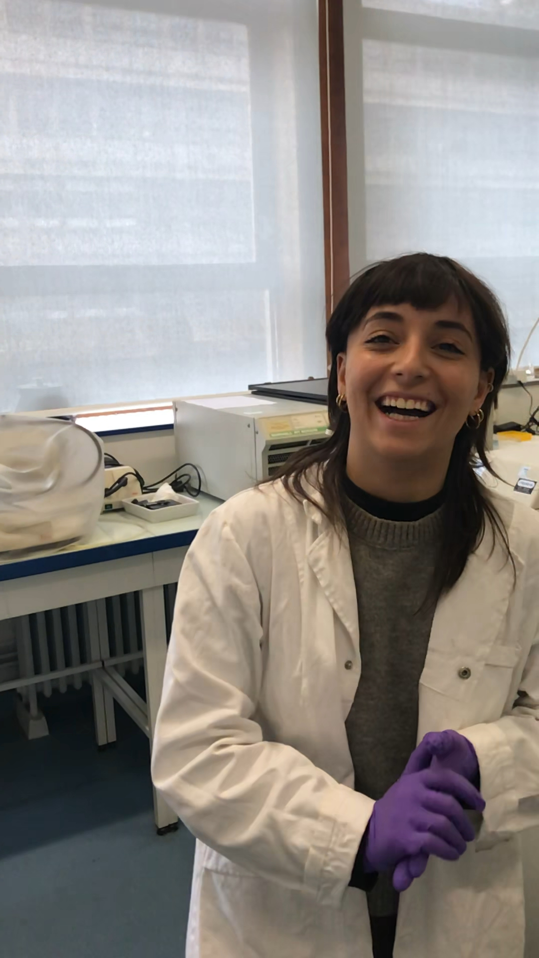
This audiovisual piece emerged from a collaboration with the GlioME team, from research they generously shared, and from on-site recordings they made with me during a visit to their glioblastoma laboratory. They later sent microscopy videos, images, and datasets, to which I replied with questions. What began as an

invitation simply to “listen” gradually became an attempt to let something of the complexity and fragility of their work be felt through sound and image. The name GlioME—gliomagenesis and microenvironment—stayed with me as a kind of thread: while their research traces the tumour’s microenvironment, my position attempts to listen to the environment and process that encompasses their work.
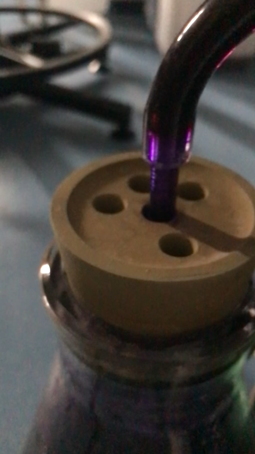
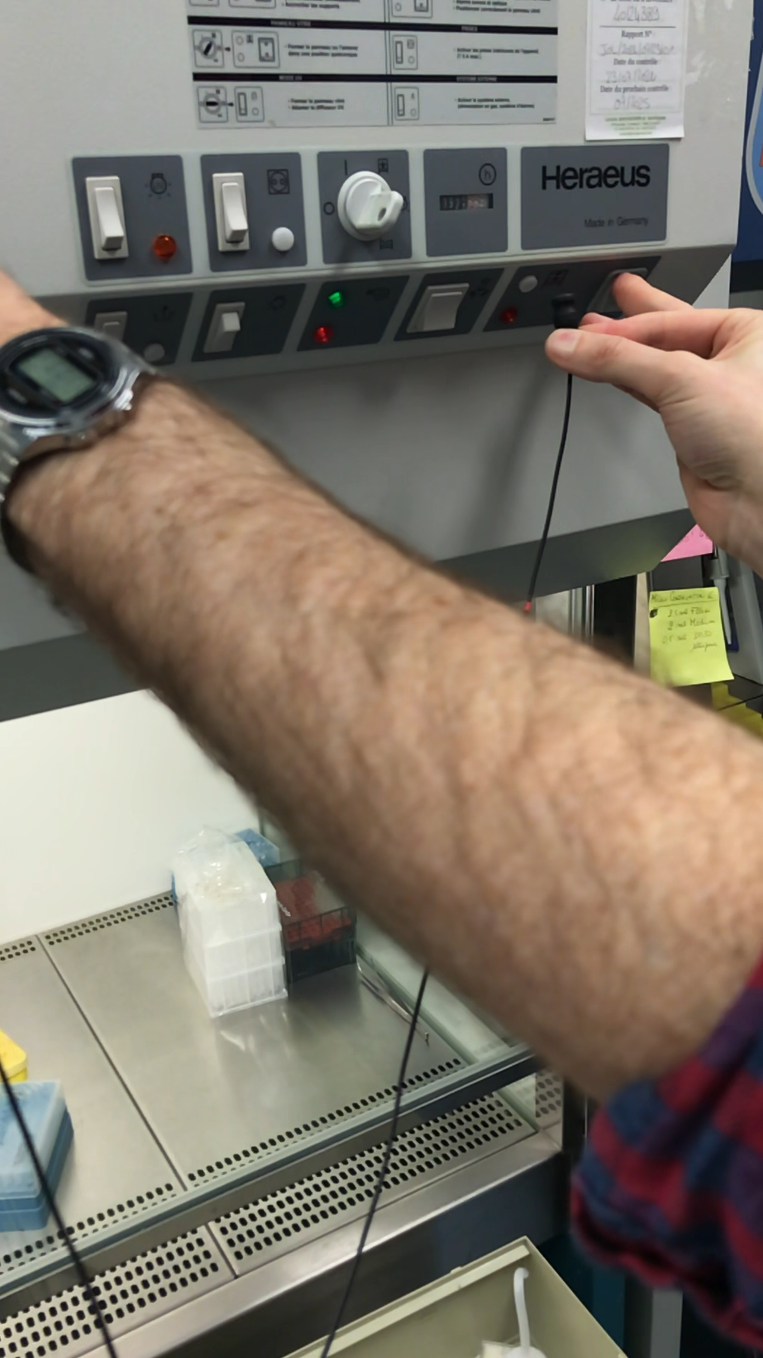
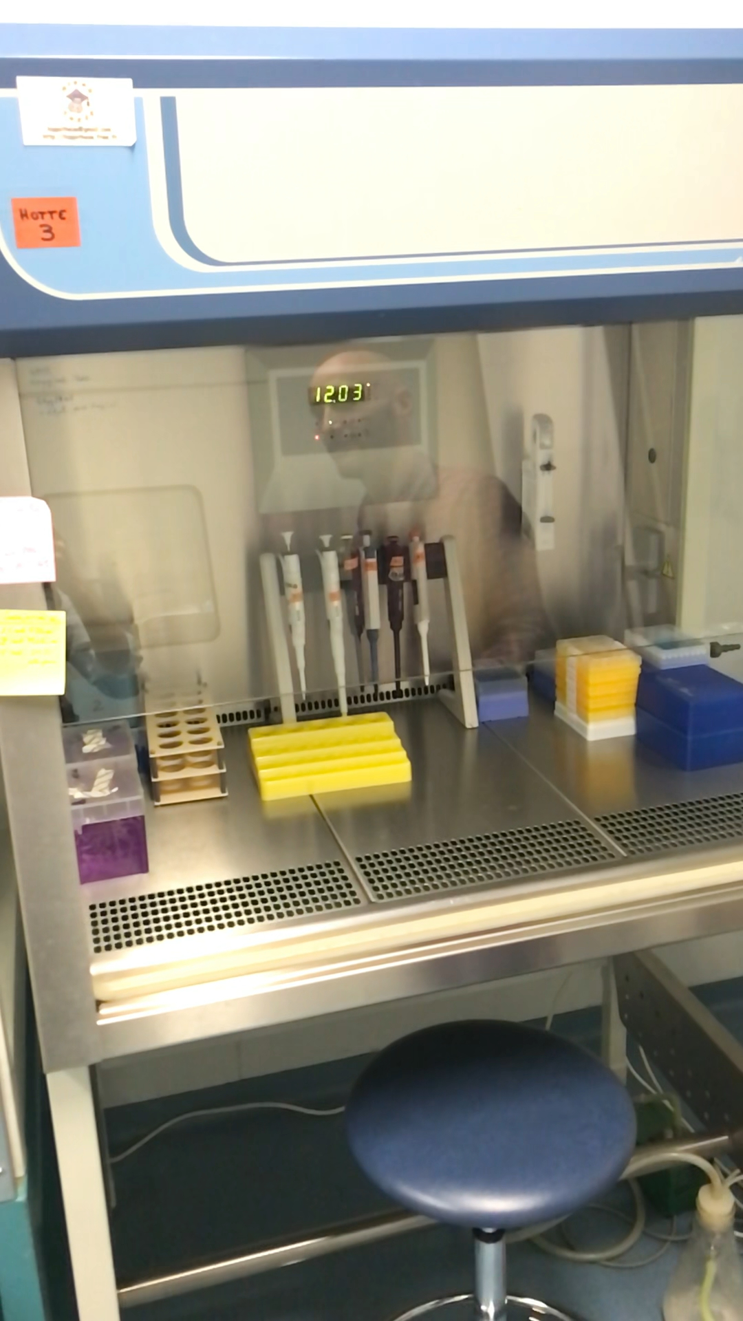
The material I worked with was both technical and personal. On one side were 3D brain “voyages” showing immunostained vasculature and immune infiltration; microscopy images; and spreadsheets describing changing conditions, distances between cells, vessels and tumours, and moments of contact between microglia and lymphocytes. On the other side were the intricate, percussive sounds and gestures of machines in the lab, and the voices of the researchers as they spoke about their practice. Nathalie Baeza-Kallée, Raphaël Berges, Nora Essakhi, Emmanuel Snacel-Fazy and Roberta Stacchini each offered on-camera time; their descriptions of motivations, constraints and long arcs of experimentation shaped how I understood the data, and the care with which I aimed to handle it. I was also struck by their own playful curiosity about sound and music—in their comments, in what they listened to, and in the way they leaned in to hear the lab with me—which fed back into how I explored the recordings and sculpted the piece.

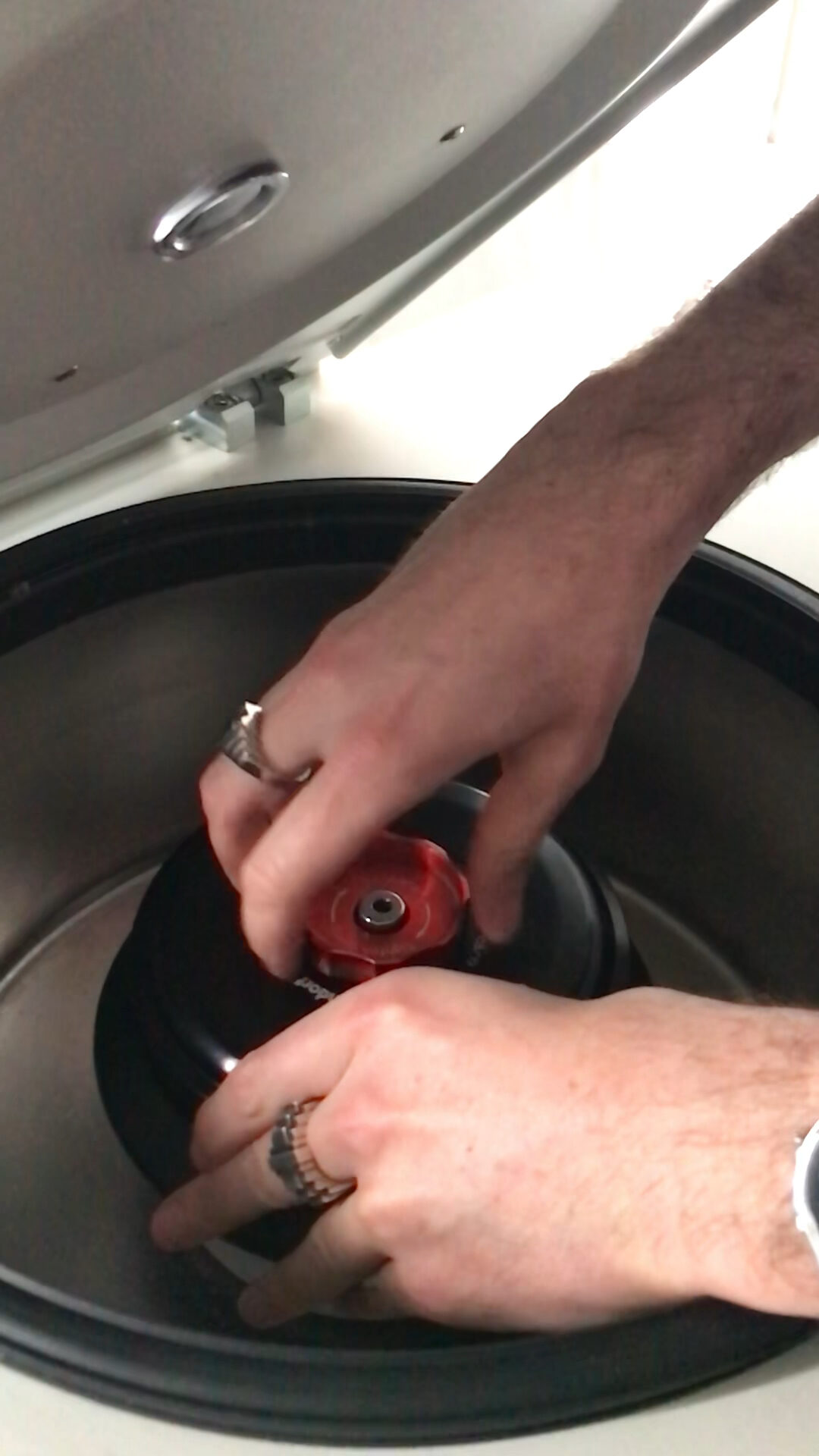
In developing the work I treated the lab recordings as instruments and the datasets as ways of shaping them, rather than as straightforward illustrations. The resonance of tubes became a kind of bass; the cyclical motion of magnetic shaker-stirrers stretched into pads; pipette releases turned into percussive
articulations; and the small mechanical turns of centrifuge caps and rotors became scratch-like textures. These sounds were modulated by numerical patterns extracted from the spreadsheets: changes in cell counts, distances to vessels or tumours, or inferred contact events subtly thickened or thinned the sound field, altered brightness and density, or shifted spatialisation. In parallel, the microscopy videos were edited, looped and placed in relation to these structures so that their movements – rotations, fly-throughs, the gradual appearance of green within red – could echo or counterpoint the temporal patterns derived from the data.
Spending time with their measurements and images made the sophistication, ingenuity and persistence of their research palpable: the steps between a living brain, a stained section, an image stack, an analysis script, and finally a figure or table.
Aurélie Tchoghandjian’s welcoming invitation, and the entire team’s openness – allowing me to see and hear their processes, to ask naïve questions, and to take time exploring connections – shaped the tone(s) of the piece. Rather than trying to explain glioblastoma or translate their findings into a single message, the work invites a shared space of attention where cells, instruments, images, voices and numbers coexist, sometimes clearly, sometimes obliquely. It is intended as a gesture of respect and curiosity toward the forms of care, rigour and imagination that sustain their practice.
Sources
Scientific data & imagery
- Tchoghandjian, Aurélie (2023). Sphericite & Area related to vessels D28 twophoton.xlsx. figshare. Dataset. https://doi.org/10.6084/m9.figshare.24679695.v1
- Tchoghandjian, Aurélie (2023). Distance TMEM119 – vessels 4X light sheet.xlsx. figshare. Dataset. https://doi.org/10.6084/m9.figshare.24679689.v1
- Derived analysis tables
Internal Excel compilation “Résultats pour sonification.xlsx” prepared by the GlioME team (Aix-Marseille Université / AP-HM), containing derived measures such as “Cells to Tumor”, “Cells to Vessel”, and “Contact microg-lymphocytes” for control vs treated conditions. Used with permission as the basis for the sonification score. - 3D brain ‘voyage’ videos and microscopy sequences
Immunostained 3D reconstructions of vascular and immune architecture in GL261-DsRED glioma models (including Lyve1-647 vasculature channels and TMEM119/CD8⁺ labeling), provided directly by the GlioME laboratory as part of the same experimental program as the above datasets, and edited for this project.
Interviews & recorded demonstrations
Audio and video recordings made in the laboratory with GlioME team members, including:
Nathalie Baeza-Kallée, Raphaël Berges, Nora Essakhi, Emmanuel Snacel-Fazy, Roberta Stacchini
These conversations and demonstrations provided the spoken material and many of the lab-instrument sounds used in the piece.
GlioME: Gliomagenèse et MicroEnvironnement, at the Institute of Neurophysiopathology (INP), Aix-Marseille Université, Inserm, CNRS, and Hôpitaux Universitaires de Marseille (AP-HM).
- Team leaders: Emeline Tabouret & Aurélie Tchoghandjian
- Researchers / faculty: Chiara Bastiancich, Olivier Chinot, Dominique Figarella-Branger, Laëtitia Padovani, Romain Appay, Corinne Bouvier, Alessandra Pagano-Aurrand Lions, Emmanuel Snacel-Fazy.
- Engineers / Technical staff: Carole Colin, Carine Jiguet-Jiglaire, Nathalie Baeza-Kallée, Raphaël Berges, Abigaëlle Gros, Aurélie Soubéran, Soline Toutain, Philippe Morando, Lënaig Bakar.
- PhD students: Alexandre Bertucci, Roberta Stacchini, Nora Essakhi.